When Should You Start Getting Breast Exams?
The words “breast cancer” trigger concern among women, as most know someone who has undergone treatment.
Finding breast cancer early is important in preventing deaths from breast cancer. When breast cancer is discovered when it’s small, at an early stage, treatment can be successful.
Doctors tell us it’s critical to have regular breast exams to ensure the best outcome. This is the most reliable way to find breast cancer early.
Every woman should have a clinical breast exam every one to three years starting at age 20. This should continue every year starting at age 40. If you have a strong family history of breast cancer, a clinical breast exam may be recommended more frequently.
If you are age 50 or over, your breast exam will include a physical exam as well as a mammogram.
Also, women with a strong family history of breast cancer should consider speaking with a genetic counselor.
Making Your Appointment for a Breast Exam
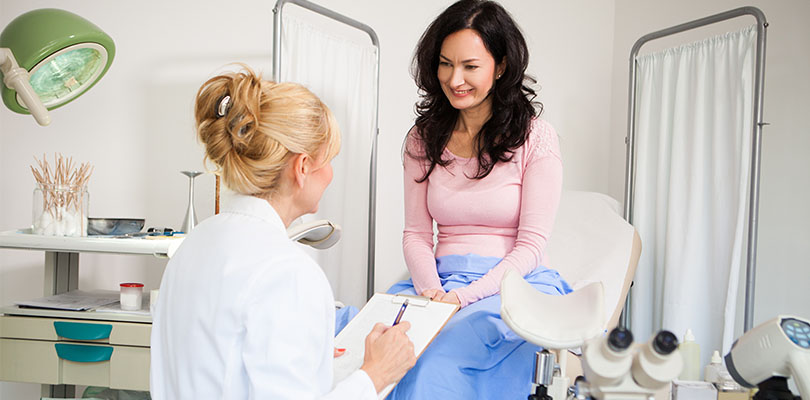
A breast exam is conducted by a health professional (your doctor, nurse, nurse practitioner, or physician assistant). This is conducted as an important part of your routine checkup.
Doctors advise timing your appointment soon after your menstrual period ends to ensure an optimal exam. Your breasts won’t be as tender and swollen, which makes it easier to detect any unusual changes.
What Happens During a Breast Exam?
First, your health care provider will ask detailed questions about your health history, including your menstrual and pregnancy history. You will be asked the age when you started menstruating. If you have children, how old you were when your first child was born?
For the exam, you undress from the waist up. Your health care provider will look at your breasts for changes in size, shape, or symmetry.
Your provider may ask you to lift your arms over your head, put your hands on your hips or lean forward. He or she will examine your breasts for any skin changes including rashes, dimpling, or redness. The area under both arms will also be examined.
Your health care provider will gently press around your nipple to check for any discharge. If there is discharge, a sample may be collected for examination under a microscope.
When Should I Start Getting Mammograms?
The United States Preventive Services Task Force (USPSTF) recommends:
- Women ages 50 to 74 years should get a mammogram every two years.
- Women younger than age 50 should talk to a doctor about when to start and how often to have a mammogram.
What Is a Mammogram?
A mammogram is a low-dose x-ray exam of the breasts to look for changes that are not normal. A mammogram allows the doctor to have a closer look for changes in breast tissue that cannot be felt during a breast exam.
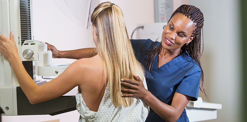
You stand in front of a special x-ray machine. The radiologic technician is the person who takes the x-rays – placing your breasts, one at a time, between an x-ray plate and a plastic plate.
These plates are attached to the x-ray machine and compress the breasts to flatten them. This spreads the breast tissue out to obtain a clearer picture.
‘Anxiety’ is often used to apply to mulitiple disorders and as an umbrella diagnosis. Start on the path to proper treatment by understanding anxiety today.
You may be a little uncomfortable during this process. You will feel pressure on your breast for a few seconds. This feeling only lasts for a few seconds, and the flatter your breast, the better the picture. Most often, two pictures are taken of each breast — one from the side and one from above.
A screening mammogram takes about 20 minutes from start to finish.
It’s worth the discomfort. Mammography can frequently detect the disease in its early stages, often before it can be felt during a breast exam. Research has shown that mammography can increase breast cancer survival.
What if My Screening Mammogram Shows a Problem?
Take heart that most abnormalities on a mammogram are NOT breast cancer.
The small group of women with abnormalities need further evaluation such as diagnostic mammography, breast ultrasound, or needle biopsy.
How Does an Abnormality Appear on a Mammogram?
An abnormality might be called a nodule, mass, lump, density, or distortion:
- A mass (lump) with a smooth, well-defined border is often benign. Ultrasound is needed to see and describe the inside of a mass. If the mass contains fluid, it is called a cyst.
- A mass (lump) with an irregular border or a starburst appearance might be cancerous, and a biopsy is usually recommended.
- Microcalcifications (small deposits of calcium) are another type of abnormality. They can be classified as benign, suspicious, or indeterminate.
- Most microcalcifications are benign. Depending on how the microcalcifications appear on the additional studies (magnification views), a biopsy might be recommended.
After this additional evaluation is complete, most women who have potential abnormalities on a screening mammogram are found to have nothing wrong.
What to Know if the Abnormality Looks Like Cancer
If you have a screening test result that suggests cancer, your doctor must find out whether it is indeed cancer or some other cause. Your doctor will ask about your personal and family medical history.
Your doctor also may order some of these tests:
- Diagnostic mammogram to focus on a specific area of the breast
- Ultrasound, an imaging test that involves sound waves to create a picture of your breast. The images may show whether a lump is solid or filled with fluid.
- A cyst is a fluid-filled sac. Cysts are not cancer. But a solid mass may be cancer.
- After the test, your doctor can store the pictures on video or print them out. This exam may be used along with a mammogram.
- Magnetic resonance imaging (MRI), which uses a powerful magnet linked to a computer. MRI makes detailed pictures of breast tissue. Your doctor can view these pictures on a monitor or print them on film. MRI may be used along with a mammogram.
- Biopsy, a test in which fluid or tissue is removed from your breast to help find out if there is cancer. Your doctor may refer you to a surgeon or to a doctor who is an expert in breast disease for a biopsy.
Next Steps to Take: Getting a Second Opinion
Your doctor is the best source of information at every step. Make sure you ask every question you have.
You might also consider getting a second opinion from another cancer specialist. Talk to your first doctor about this. Your primary doctor will not be offended; they respect your right to get all the information you need to move forward.
Depending on your situation, a second opinion can change the details of your diagnosis, suggest other treatment options, or even change the course of treatment entirely.
For example, patients must submit all their lab work, including pathology reports, x-rays and other test results. They will want to know two things: whether or not they have the correct diagnosis, and if they are on the optimum treatment plan.
Ask your health insurance company if it covers a second opinion. You can see another breast cancer specialist in your city.
Also, if you’re seeing a second doctor in person, you must have your first-opinion records sent ahead to the second doctor.
The Takeaway
The bottom line is this – you want peace of mind that you have done everything you could. Don’t let this process drag into a wild goose chase of wishful thinking.
It’s best to get your expert opinions, then make a decision. Don’t waste time - as early treatment ensures the very best outcome.


